Hey PS+R Crew!
We’ve got BIG news — PS+R has moved! 🎉
We are beyond grateful for our first home at Stone Climbing. Their support was instrumental in bringing the PS+R dream to life, and they will always remain a valued partner. While we’ll miss the daily energy and positive vibes of the Stone Climbing crew, we’re excited about the possibilities this next chapter brings.
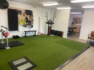
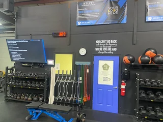
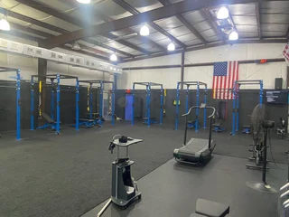
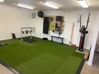
As of Monday, August 11th, PS+R officially relocated to Ktlyst Fitness!
📍 101 Liberty Center Pl, St. Augustine, FL 32092 (just west of SR 16 and I-95).
Ktlyst has been a trusted partner over the past year, offering two expansive strength and conditioning spaces where our athletes thrive under the guidance of expert coaches. This move allows us to deepen that partnership and provides the physical space needed to expand our expertise in return-to-sport training.
Come check out our new facility and see what the PS+R and Ktlyst partnership has to offer!
Share the love!
Share the love and leave us a review on our google business page: https://g.page/r/CXZ2JA04uELUEAE/review

Welcome Dr. Trent Salo
This month, PS+R is thrilled to welcome Dr. Trent Salo to the team. Trent will be a PS+R twice a month performing ultrasound tissue characterization- a type of advanced clinical ultrasound- for the evaluation and management of chronic tendon pain. Read on below to learn more about our collaboration with Dr. Salo and the tendon lab and find out how this unique, world-class service might help your performance goals right here at home.
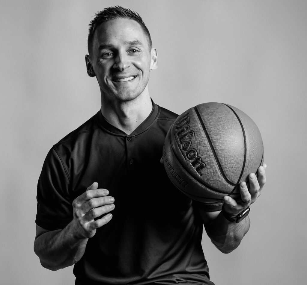
If you’d like to schedule a time with Trent, reach out to him with his email below, or to our team to help direct you!
Trent’s email: Trent@performancesciencerehab.com

Ultrasound Tissue Characterization: Advanced Imaging for Tendon Health Assessment
Imagine being able to see inside your tendons with remarkable precision in just 45 seconds—Ultrasound Tissue Characterization (UTC) makes this possible through a completely painless process. At the push of a button, an ultrasound transducer automatically collects 600 successive images, capturing data every 0.2mm throughout the entire length of a tendon. Sophisticated computer algorithms then analyze this comprehensive dataset to identify and quantify tendon structure with unprecedented detail, providing intuitive color-coding that instantly reveals tissue condition: green represents intact, well-aligned fibers indicative of healthy tendon; blue shows discontinuous but organized fibers; red indicates fibrillar tissue with some disorganization; and black represents compromised amorphous tissue. This objective, standardized assessment transforms what was once subjective visual interpretation into precise, quantifiable data that can be reliably tracked and monitored over time.
The clinical applications of UTC have revolutionized both preventive care and active treatment guidance, fundamentally changing how we approach tendon health management. While early research by Cook et al. and Docking et al. showed conflicting relationships between tendon structure and pain, recent evidence has supported UTC’s clinical significance. Multi-institutional studies by Cushman et al. demonstrate that ultrasound screening effectively predicts injury risk in collegiate athletes, while research by Rabello et al. confirms that clinical improvements directly correlate with structural tendon changes visible on imaging. For patients experiencing tendon pain, UTC reveals the specific location, distribution, and severity of tissue changes, enabling targeted treatment strategies rather than generic rehabilitation protocols. In my practice, UTC provides repeatable, objective evidence that guides personalized intervention strategies—helping patients understand not just that they have tendon pain, but exactly what’s happening within their tendon structure and how to optimize their path to recovery.
References:
- Cook, J. L., et al. (2000). Prospective imaging study of asymptomatic patellar tendinopathy in elite junior basketball players. Journal of Ultrasound in Medicine, 19(7), 473-479.
- Corrigan, P., et al. (2020). Tendon Morphology and Mechanical Properties Are Associated With the Recovery of Symptoms and Function in Patients With Achilles Tendinopathy. Orthopaedic Journal of Sports Medicine, 8(5).
- Cushman, D. M., et al. (2024). Ultrasound Screening of Achilles Tendon, Patellar Tendon, and Plantar Fascia for Pain Development in Collegiate Athletes. Orthopaedic Journal of Sports Medicine, 12(11).
- Cushman, D. M., et al. (2025). Ultrasound as a predictor of time-loss injury for the patellar tendon, Achilles tendon and plantar fascia in division I collegiate athletes. British Journal of Sports Medicine.
- Cushman, D. M., et al. (2024). Sonographic Assessment of Asymptomatic Patellar and Achilles Tendons to Predict Future Pain: A Systematic Review and Meta-analysis. Clinical Journal of Sport Medicine.
- Docking, S. I., et al. (2015). Tendinopathy: Is imaging telling us the entire story? Journal of Orthopaedic and Sports Physical Therapy, 45(11), 842-852.
- Rabello, L. M., et al. (2020). Association between clinical and imaging outcomes after therapeutic loading exercise in patients diagnosed with achilles or patellar tendinopathy. Clinical Journal of Sport Medicine, 30(4), 390-403.
PS+R In The News | September 2025
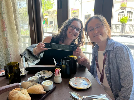
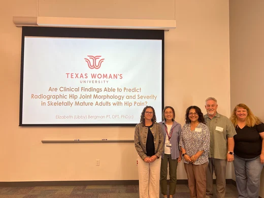
Libby’s Research
Libby successfully defended her dissertation this month which marks her near completion of her PhD. She is finalizing the format of her dissertation and will graduate in December, 2025.
Where in the world is Steve and the USMNT?
Steve and the USMNT have two matches for this upcoming October Window as preparations for the World Cup in the summer of 2026 heat up. Matches will be played in Austin Texas to play Ecuador on October 10th and Denver Colorado on October 14th to face off against Australia. It is hard to believe that we are only 8 matches away from hosting the World Cup in 2026!
Thanks for staying connected with us! See you around!!